Medical Image Computing
Computing for
Diagnosis and TherapyAdapted from an article requested by Postgraduate UK.
Within a few months of the discovery of X-rays by Wilhelm Röntgen in 1895, X-ray images were used to plan and guide a surgical intervention. In the field of medical imaging, the themes of innovation in diagnosis and treatment have continued over the past century and the UK has gained two Nobel prize winners; Godfrey Hounsfield for the development of computer assisted tomography (CT) and Peter Mansfield for discoveries concerning magnetic resonance imaging (MRI). Hounsfield’s work used data from X-rays taken at different angles to perform tomography – the calculation of image slices through the body. MRI also produces three dimensional data but is based on magnetic fields and radio waves rather than X-rays. MRI and CT are complementary - each providing images sensitive to different types of tissue.
In addition to MRI and CT, we also have ultrasound imaging based on the reflection of sound waves and positron emission tomography (PET) and nuclear medicine that detect the decay of radio-isotopes. These isotopes are added to compounds that concentrate at sites of specific tissues or diseases enabling them to be visualized in imaging. Furthermore, medical images come from surgical microscopes, endoscopic examinations, photographs and video as well as emerging technologies such as optical tomography whereby light is shone through the head at multiple angles. This wealth of data brings new information and great opportunities to apply computing methods for enhancing diagnosis, helping plan, guide and assess therapies and for the investigation of fundamental questions about the working of the human body.
Computers in Imaging
Acquisition systems
produce digital data that is converted to images and ultimately viewed
by
radiologists and surgeons. Present throughout this chain are computers,
not
just as convenient tools for storage, workflow and display, but also in
the
control of the scanning, the alignment of images, detection of
different tissue
types, calculation of quantitative measures, generation of new images
such as
maps of active regions in the brain and surgical guidance. Increasingly
the
hardware of scanners is under software control and we have the
opportunity to
produce algorithms that will intelligently enhance the image
acquisition by
responding in real time to patient data.
A
key tool
for generating new information is image registration, a field in which
the UK
has been very active in research. Registration algorithms compute the
transformation needed to align, or warp, one image into another. With
the
ability to align images, comparisons between images taken on different
days can
reveal small changes in tissue size, for example the slow shrinkage of
the
brain over many years due to dementia. Finding the transformation from
one
image to another can be used to measure motion, for example a cine
series of
cardiac images can show abnormalities in heart wall motion due to
tissue
damaged by a heart attack. In another application of registration,
images from
different patients are aligned into one common space. A representative
image or
atlas can then be computed, against which new patients can be compared.
Registration is used in the fusion of data from different scanners
leading to
enhanced understanding, for example, a PET image is sensitive to the
uptake of
glucose by tumours and an MRI image can show other anatomy in detail -
combining the two provides a richer source of information to guide
patient
care.
Obtaining quantitative measures is vital for enabling patient information to be compared with existing knowledge or to assemble new measures of anatomy and physiology. UK researchers have developed methods for modelling shape to study structural and functional variation in health and diseased states. Research is also using shape information to find organs and bones in images and to guide surgery.
In surgery and interventions where a catheter is inserted through a blood vessel into the heart, images from previous scans can be used to help guide the operator during the procedure. Registration techniques, and the 3D tracking of equipment, are used to visually present images taken prior to the procedure, which are overlaid on the new surgical scene. The world’s first MRI guided cardiac catheter intervention took place recently at Guy’s Hospital, King’s College London.
Patient movement resulting from involuntary motion, cardiac pulsation, respiration and flowing blood can all blur MR images. Novel algorithms for correcting images are being developed at Imperial College London and UCL to aid diagnosis.
Medical and Technological Drivers
Faster scans at higher resolution and with better image quality are always in demand. Our enhanced understanding of the genome is driving a desire to observe changes at the molecular scale whilst wanting to consider the patient as a whole. A dream goal might be the biological equivalent of Google Earth that enabled zooming from a whole body image down to the cellular and then molecular levels.
Detector technology is improving and providing ever more data. In MRI, the numbers of coils used to receive the signal has increased by an order of magnitude. In CT, the detectors that acquire the X-ray signal at each angle now have many more elements, generating much more data. In PET imaging, modern scanners are now combined with a CT scanner and in ultrasound, micro bubbles injected as contrast provide harmonic signals in addition to the main signal.
These large quantities of data present challenges for computer algorithms and are very demanding of memory requirements. The increased availability of 64-bit machines and the ability to connect large numbers of PCs to form a cluster are addressing these issues. For example, the CMIC group at UCL have their own 60 node, 64-bit cluster and there are e-science projects that use a national grid of computers. One e-science project called IXI is collecting brain images from 600 people at three different sites. The aim is for a doctor to be able to see at a glance the normal range of sizes and shapes of each brain structure, overlaid on the patient’s own scan, assisting diagnosis. To provide this information, the grid will access data that may be stored in distributed places and perform the necessary calculations on machines located anywhere on the grid network. Just like the electricity grid, the user draws resources without concern for where they are generated.
Imaging in clinical trials
Thriving Industry
Postgraduate Opportunities
In
2007, UCL plans to start an MSc dedicated to Medical Image Computing.
The course can be taken full or part time and scholarships are
available to some
students.
The
UK
Engineering and Physical Sciences Research Council recognised the
strength of
medical imaging by funding a six-year project that now links Imperial
College
London, Kings College London, Manchester, Oxford and UCL. These groups,
and
others in the country, provide opportunities for obtaining a PhD in
medical
image computing. A bi-annual summer school brings together an
international
teaching faculty to provide lectures and workshops for Postgraduates
studying
in the UK and overseas.
Summary
Within
the
UK, university research in medical image computing is well-funded,
industrial
activity ranges from start-up companies to global pharmaceutical
organisations
and there is substantial investment by the government in the healthcare
sector.
This creates a healthy environment for Postgraduate study and research
and in a
subject that brings together computing, medicine, healthcare, biology,
maths,
engineering and physics for applications that benefit healthcare and
well-being.
 |
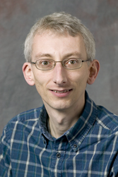
David Atkinson is a lecturer in the Centre for Medical Image Computing at University College London. Since 1996 he has been researching novel algorithms to improve magnetic resonance images. He is currently preparing a new MSc in Medical Image Computing at UCL for launch in 2007. |